In the early course of the disease, clinical diagnosis of mvd is difficult to distinguish from other tropical febrile illnesses, because of. The marburg virus does not contain the polyadenylation sequence that is found in the ebola gp gene. Check spelling or type a new query. Molecular structures and visual images of the proteins of marburg virus are essential for the development of antiviral drugs. Johnson ed, johnson bk, silverstein d, et al. A cousin virus of ebola. Cases in marburg (germany) and belgrade in 1967 led to the initial recognition of the disease. This means that both have the shape of a wire, and therefore the same structure. The marburg virus contains seven structural proteins. Viewed under electron microscopy, the viruses show particles shaped like elongated filaments, sometimes coiled into strange shapes, that give the filoviridae family its name.
It is unknown how marburg virus first transmits from its animal host to humans; The virus was discovered in 1967 in germany, when monkeys imported to the institute of virology in marburg infected researchers. Johnson ed, johnson bk, silverstein d, et al. In addition, exposure to an infected human is high risk factor.

Risk factors include exposure to african green monkeys and certain bats;
Gastrointestinal distress, including watery diarrhea, nausea, and cramping, often around three days after symptoms appear. Marburgvirus, genus of viruses in family filoviridae, known for causing severe disease in humans and other primates.one species has been described, marburg marburgvirus (formerly lake victoria marburgvirus), which is represented by two viruses, ravn virus (ravv) and marburg virus (marv). Marburg virus is a member of the filoviridae family, and is an elongated filamentous molecule, highly variable in length, but typically around 1000 nm long with a uniform diameter of 80 nm (2,3). Suivez l'évolution de l'épidémie de coronavirus / covid19 dans le monde. One key protein in the marburg virus life cycle is … For the marburg virus to infect the host's cell an essential element is needed. The marburg virus is from the same family as ebola: Check spelling or type a new query. Risk factors include exposure to african green monkeys and certain bats; They were due to laboratory work using african green monkeys. Crystal structure of the marburg virus nucleoprotein core domain chaperoned by a vp35 peptide reveals a conserved drug target for filovirus j virol. It is unknown how marburg virus first transmits from its animal host to humans; Associated diseases hemorragic fever, often fatal. The virus is considered to be extremely dangerous.
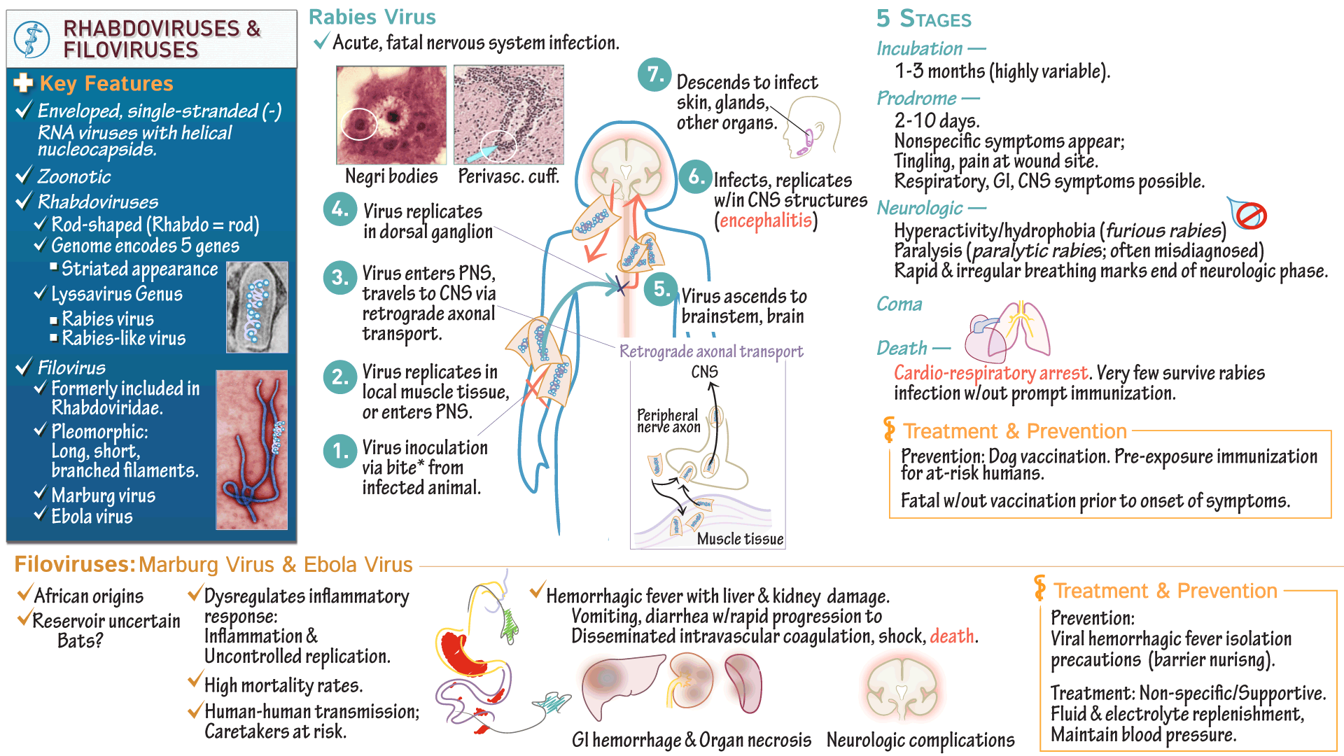
Marburg haemorrhagic fever is a severe and highly fatal disease caused by a virus from the same family as the one that causes ebola haemorrhagic fever. The marburg virus is from the same family as ebola: Viewed under electron microscopy, the viruses show particles shaped like elongated filaments, sometimes coiled into strange shapes, that give the filoviridae family its name. Marburg virus is a member of the filoviridae family, and is an elongated filamentous molecule, highly variable in length, but typically around 1000 nm long with a uniform diameter of 80 nm (2,3). Suivez l'évolution de l'épidémie de coronavirus / covid19 dans le monde. Like ebola, marburg virus disease can cause severe hemorrhaging that leads to shock, organ failure, or death. Marburg virus structure and transmission. Marburg virus disease (mvd), formerly known as marburg haemorrhagic fever, is a severe, often fatal illness in humans. Crystal structure of the marburg virus nucleoprotein core domain chaperoned by a vp35 peptide reveals a conserved drug target for filovirus j virol. East africa, kenya, congo and angola.

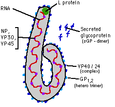
As you can see in figure 2, the green helical structure is the genomic rna surrounded by polymer of nucleoproteins (np).
This means that both have the shape of a wire, and therefore the same structure. Smith dh, johnson bk, isaacson m, et al. The proteins of ebola and marburg are likewise similar. Molecular structures and visual images of the proteins of marburg virus are essential for the development of antiviral drugs. The world health organization (who) rates it as a risk group 4 pathogen. In humans, marburgviruses are responsible for marburg virus disease (mvd), a zoonotic disease that is. Marburg virus structure and transmission. Marburg virus, a cousin of ebola virus, causes severe hemorrhagic fever, with up to 90% lethality seen in recent outbreaks. (d) topology diagram of marburg virus vp24, with secondary structure elements sequentially numbered and colored from n to c as described for panels a to c. Marburg virus disease (mvd), formerly known as marburg haemorrhagic fever, is a severe, often fatal illness in humans. Check spelling or type a new query. In the early course of the disease, clinical diagnosis of mvd is difficult to distinguish from other tropical febrile illnesses, because of. Marburg virus diagram / research muhlberger lab.
Johnson ed, johnson bk, silverstein d, et al. In the early course of the disease, clinical diagnosis of mvd is difficult to distinguish from other tropical febrile illnesses, because of the similarities in the clinical symptoms. Characterization of a new marburg virus isolated from a 1987 fatal case in kenya. The virus was discovered in 1967 in germany, when monkeys imported to the institute of virology in marburg infected researchers. Analyzing such images sheds light on the structure of the virus and the mechanisms by which it is assembled. Marburg virus structure and transmission. In the early course of the disease, clinical diagnosis of mvd is difficult to distinguish from other tropical febrile illnesses, because of. Marburg virus, a cousin of ebola virus, causes severe hemorrhagic fever, with up to 90% lethality seen in recent outbreaks. As with most of griyo's viruses, marburg is named for a human/animal virus.
Marburg and ebola viruses are both members of the filoviridae family (filovirus).
Marburg virus disease (mvd), formerly known as marburg haemorrhagic fever, is a severe, often fatal illness in humans. Marburg virus diagram / research muhlberger lab. As you can see in figure 2, the green helical structure is the genomic rna surrounded by polymer of nucleoproteins (np). Marburg virus is a member of the filoviridae family, and is an elongated filamentous molecule, highly variable in length, but typically around 1000 nm long with a uniform diameter of 80 nm (2,3). The proteins of ebola and marburg are likewise similar. Marburg virus is the causative agent of marburg virus disease (mvd), a disease with a case fatality ratio of up to 88%. Marburgvirus, genus of viruses in family filoviridae, known for causing severe disease in humans and other primates.one species has been described, marburg marburgvirus (formerly lake victoria marburgvirus), which is represented by two viruses, ravn virus (ravv) and marburg virus (marv). In the early course of the disease, clinical diagnosis of mvd is difficult to distinguish from other tropical febrile illnesses, because of. The virus is considered to be extremely dangerous. Marburg virus structure and transmission. In humans, marburgviruses are responsible for marburg virus disease (mvd), a zoonotic disease that is. As with most of griyo's viruses, marburg is named for a human/animal virus.
Johnson ed, johnson bk, silverstein d, et al marburg-virus. Marburg virus disease was initially detected in 1967 after simultaneous outbreaks in marburg and frankfurt in germany;

Gastrointestinal distress, including watery diarrhea, nausea, and cramping, often around three days after symptoms appear.

(d) topology diagram of marburg virus vp24, with secondary structure elements sequentially numbered and colored from n to c as described for panels a to c.

In addition, exposure to an infected human is high risk factor.

Marburg and ebola viruses are both members of the filoviridae family (filovirus).

Marburg virus was first recognized in 1967, when outbreaks of hemorrhagic fever occurred simultaneously in laboratories in marburg and frankfurt, germany and in belgrade, yugoslavia (now serbia).

The marburg virus does not contain the polyadenylation sequence that is found in the ebola gp gene.

Marburg virus is the causative agent of marburg virus disease (mvd), a disease with a case fatality ratio of up to 88%.

Cases in marburg (germany) and belgrade in 1967 led to the initial recognition of the disease.

Crystal structure of the marburg virus nucleoprotein core domain chaperoned by a vp35 peptide reveals a conserved drug target for filovirus j virol.

Marburgvirus, genus of viruses in family filoviridae, known for causing severe disease in humans and other primates.one species has been described, marburg marburgvirus (formerly lake victoria marburgvirus), which is represented by two viruses, ravn virus (ravv) and marburg virus (marv).

A cousin virus of ebola.

The virus is considered to be extremely dangerous.

The marburg virus does not contain the polyadenylation sequence that is found in the ebola gp gene.

Marburg virus causes symptoms that come on suddenly and become increasingly severe.

Marburgvirus, genus of viruses in family filoviridae, known for causing severe disease in humans and other primates.one species has been described, marburg marburgvirus (formerly lake victoria marburgvirus), which is represented by two viruses, ravn virus (ravv) and marburg virus (marv).

Crystal structure of the marburg virus nucleoprotein core domain chaperoned by a vp35 peptide reveals a conserved drug target for filovirus j virol.

Marburg virus disease is caused by viruses that produce symptoms of fever, chills, headaches and muscle aches early in the disease;

Comments and questions to info@ncbi.nlm.nih.gov

Gastrointestinal distress, including watery diarrhea, nausea, and cramping, often around three days after symptoms appear.

Check spelling or type a new query.

In the early course of the disease, clinical diagnosis of mvd is difficult to distinguish from other tropical febrile illnesses, because of the similarities in the clinical symptoms.

Maybe you would like to learn more about one of these?

Check spelling or type a new query.

One key protein in the marburg virus life cycle is …

The five species of ebola virus are the only other known members of the filovirus family.

Marburg virus diagram / research muhlberger lab.

As with most of griyo's viruses, marburg is named for a human/animal virus.
Check spelling or type a new query.